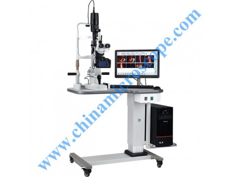
MIC-88D2 serials slit lamp microscope
Five-step drum magnification for eyepice. Sclera vein could be observed under the 40× magnification
All the optical lenses are moistureproof, mildewproof and anti-reflected ested
Large spot dismeter, slit length reach to 14mm, more comprehensive view of the eye contiton
Professional collection media
The instrument SLR camera as a collection media, real-time dynamic display, clear and vivid images, fast image acquisition.
Camera Shortcut
External shooting device spotted lesions touch of a capture clear picture immediately.
Powerful image processing functions
Measure length, area, angle, grayscale, curvature, label the lesions in the picture, and add text.
High definition image
This instrument is capable of shooting photos of up to 18 million pixels, and offers up to 40×magnification, completely show the details,
to meet the demanding requirements of the medical image, enable the lesions to be show more clearly.
A diagnostic aid
Provide a variety of common eye checkup mode parameter set to improve examination efficiency.
With medical records management and output.
Case report can be saved, manage, print.
Configuration:
• Slit lamp
• Camera interface (splitter / adapter)
• Professional SLR camera (standard configuration does not contain a camera lens)
• Bolan slit lamp image processing systerm
• Computer (optional)
• High-resolution ink-jet printer (optional)

